Search

Nuclear Medicine- Bone Scan

A nuclear medicine bone scan study involves injecting a small amount of a radioactive substance, called a radiopharmaceutical, into one of your veins. This substance travels through your bloodstream and attaches mostly to your bones. A special camera, known as a gamma camera, detects how your bones are functioning based on this activity. The radioisotope used is a very small amount of a radioactive material therefore there is no need for precautions against radioactive exposure.  This material won’t harm you, nor is there any side-effects from the injection. You won’t need to limit your contact with other people, and as you hydrate and urinate for the remainder of the day, the product will leave your system.

SPECT/CT imaging combines a SPECT (Single Photon Emission Computed Tomography) scan with a low-dose CT (Computed Tomography) scan to better pinpoint any abnormal areas. The gamma camera rotates around your body for the SPECT scan. The CT part helps localize the area and can be digitally combined with the SPECT results for more precise information.

This exam is performed by an Xray technologist with special training in nuclear medicine imaging, and the results from the scan are interpreted by a radiologist.

Types of Nuclear Medicine Scans

At Beam Radiology we offer the nuclear medicine bone scan. This can be targeted to any area of interest based on your referrer’s question or concern.

FAQs

What conditions are nuclear medicine bone scans able to detect?
  • Detects smaller hard to see fractures that Xray may not be able to catch 
  • Cancer
  • Disease
  • Trauma
  • Soft tissue injuries
What nuclear medicine exams are performed at Beam?

Bone Scans (any area of interest as requested by your care provider, or recommended by a radiologist based on the history)

How Do I prepare for my exam?
  • Tell your technologist if you are pregnant, think you might be pregnant, or are nursing. If you are pregnant, the scan will be postponed. If you might be pregnant, a blood test may be needed to confirm pregnancy status before making your booking.
  • Stay hydrated by drinking 4-5 glasses of water before and during the study. You can eat and drink as usual.
  • Inform your technologist if you feel anxious or claustrophobic in tight spaces. Your doctor may prescribe medication to help.
  • Bring photo ID, your provincial health card, and your requisition form.
  • Do not bring children who need supervision.
What can I expect at my appointment?

Upon arrival, you will check in at the front reception and may be asked to fill out some forms. You will then be escorted to an area where you may need to change into a medical gown. 

There are two parts to a nuclear medicine imaging exam:

  • Part 1: A small amount of radioactive material is injected into one of your veins. This part of the study takes about 30 minutes and may involve some imaging. You can leave the clinic afterward. Your technologist will let you know your scheduled return time, usually 2-4 hours later. During this time, avoid strenuous activities or exercise. Be sure to continue your intake of fluids as directed.
  • Part 2: When you return to the clinic, please go to the bathroom and empty your bladder.  You will be asked to lay down on the exam table with the camera positioned over you. This portion of the study, including your arrival and use of the washroom will take approximately one hour. It is important that while the camera is actively imaging you try to remain still so that the best images can be obtained. Let the technologist know if you are uncomfortable in any way while lying on the bed, and they will work to help resolve this for you. We want to make you as comfortable as possible.

After the Study

  • Sometimes, additional x-rays may be needed to assist in the imaging results. If so, these will be done after the bone scan is completed, and this can take an additional, up to, 30 minutes.
  • For the rest of your day, drink plenty of water and urinate frequently to help flush the radioactive substance from your body.
What are the risks associated with my nuclear medicine bone exam?

A nuclear medicine bone scan has a low risk of side effects, but these can include injection discomfort, allergic reactions, and radiation exposure (extremely low). 

Injection discomfort 

  • As with any injection, the injection itself may cause slight pain and redness around the site
  • There may be swelling at the injection site

Allergic reactions 

  • Allergic reactions to the radiotracers are extremely rare and usually mild
  • You should tell the nuclear medicine personnel about any allergies you may have

Your Health is Important to Us

If you have any questions or would like to learn more, please
contact us. We look forward to supporting your journey to better health.

Dr. Olivier Clerk-Lamalice​

Dr. Olivier Clerk-Lamalice

MD-MSc, FRCPC, FIPP

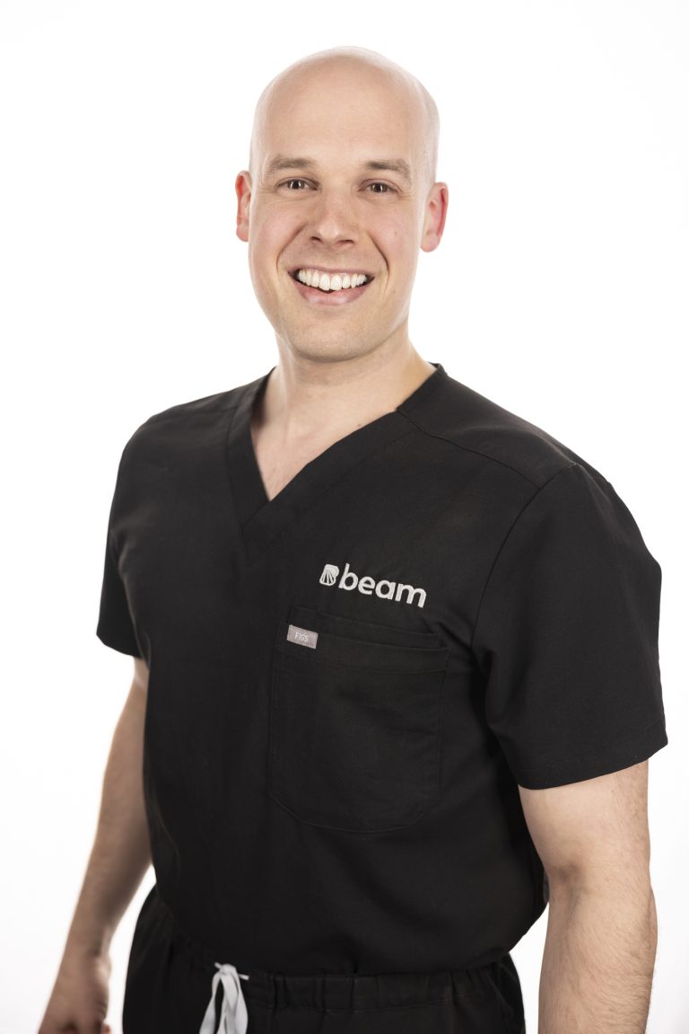
Dr. Clerk is a radiologist and fellowship-trained interventional radiologist with a wide array of experience in both interventional pain management and diagnostic imaging. In addition to providing expert patient care, Dr. Clerk places utmost importance on building a compassionate practice that recognizes patients as people, not numbers.

When you choose Beam, you can be confident that Dr. Clerk will stay with you throughout your care journey and help you make smart decisions about your pain and imaging needs.

Education & Training

Université de Sherbrooke
Medical School

Université de Sherbrooke
Residency | Diagnostic Radiology

Harvard Medical School
Fellowship | Neuroradiology

The Spine Fracture Institute
Fellowship | Interventional Pain Management