Search

CT Colonography Screening Program

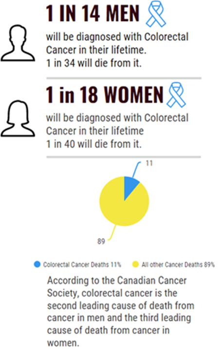
COLORECTAL CANCER IS A HIGHLY PREVENTABLE DISEASE IF IDENTIFIED EARLY

What is a CT Colonography?

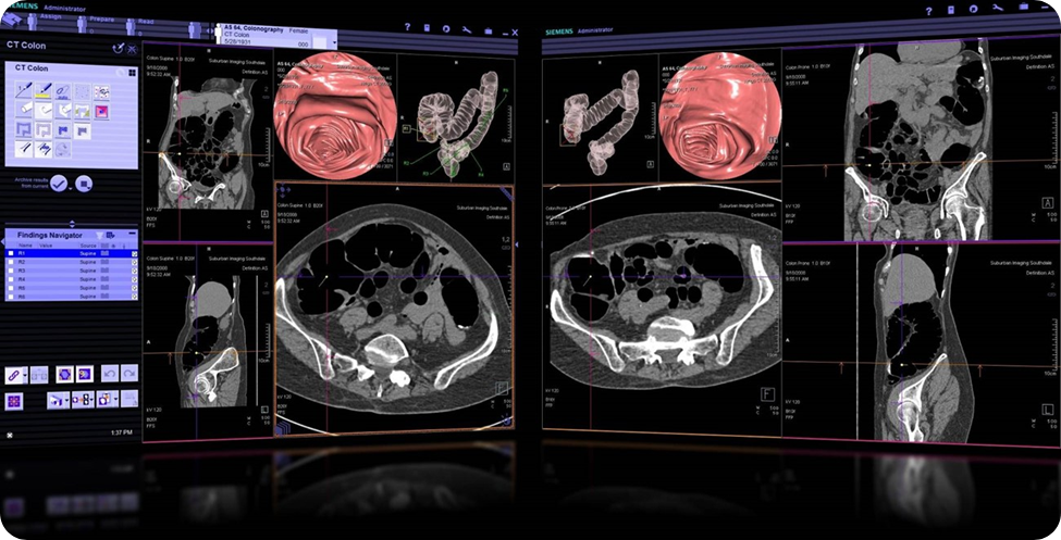
CT Colonography, also known as virtual colonoscopy, utilizes a CT scanner to provide rendered images of the entire large bowel (colon and rectum). This is done to detect polyps and masses. Colonic polyps are small growths on the lining of the colon which are a pre-cursor to cancer.

Beam - CT Scan Result

What are the Benefits?

  • Early detection of polyps or cancers allows for earlier intervention and a better prognosis
  • Colorectal cancer is a highly preventable disease if identified at the polyp stage
  • Minimally invasive and more comfortable than traditional colonoscopy
  • Faster booking and timeline for screening
  • Reduced post procedure complications

Who Should Consider This Screening Test?

  • Anyone over the age of 50 (American Cancer Society suggests age 45)
  • Have a positive FIT test (fecal immunochemical test)
  • Have a personal history of inflammatory bowel disease, colorectal cancer or radiation therapy to the abdomen or pelvis
  • Have a family history of colon cancer or polyposis
  • Have blood in the stool
  • Those who are unable to have a regular colonoscopy
CT Colonography
CT Colonography

What is Mammography?

Mammography is an essential tool for the early detection of breast cancer. Regular mammograms can help save lives by identifying breast cancer at its earliest and most treatable stage, often long before it can be felt.

Mammography is a special type of medical imaging that is used to detect and diagnose breast diseases, including breast cancer. It involves taking low-dose X-ray images of the breasts to look for any abnormalities or changes in the breast tissue even before a patient or doctor can feel a lump.

Digital Breast Tomosynthesis (DBT), also known as 3D mammography, is an advanced imaging technique that provides a three-dimensional view of the breast. It is used in combination with traditional 2D mammography to aid in the detection and diagnosis of breast cancer.

In DBT, a series of low-dose X-ray images are taken of the breast from different angles. These images are then reconstructed into a three-dimensional image of the breast, allowing radiologists to examine the breast tissue layer by layer. This provides a more detailed and accurate view of the breast, making it easier to detect small abnormalities or lesions that may be hidden or obscured in traditional 2D mammograms.

The addition of DBT to mammography has been shown to improve the accuracy of breast cancer detection, especially in women with dense breast tissue. Dense breast tissue can make it more challenging to detect cancers with conventional mammography alone, as the dense tissue can mask or hide abnormalities. DBT helps to overcome this limitation by providing clearer and more detailed images of the breast.

Why is Mammography Useful in Breast Health?

Mammography is important because it can help detect breast cancer at an early stage, when the treatment is most effective, and the prognosis is best. Regular mammograms can help identify small tumors or changes in breast tissue that may not be felt during a physical exam. By detecting breast cancer earlier, women have more treatment options available to them, often including options that are less aggressive, and a higher chance of successful treatment outcomes.

FAQs

How do I Prepare for the Appointment?
  • You will be provided with a preparation kit and instructions. This will guide you through a gentle bowel cleanse starting the day before the procedure, in the comfort of your own home. This is an important step to provide the best possible assessment of the colon. Follow the instructions provided with the kit very carefully.
  • Please arrive 30 minutes before your scheduled appointment
What Can I Expect During This Screening Test?

You will be provided with a preparation kit to guide you through a gentle bowel cleanse starting the day before the procedure, in the comfort of your own home. This is an important step to provide the best possible assessment of the colon.

On the day of the screening test:

  • Once you have checked in at the clinic, you will be shown into the changing area where you will be provided a gown and a private space to wait. 
  • Before the scan begins, you will lie on your side while a thin and flexible tube will be placed carefully into the lower colon by a trained healthcare professional. A gas will flow through this tube to gently inflate the colon. CT images will be taken while you are lying briefly on your back and then on your front. 
  • Once the images are complete, the tube will be removed.  
  • The exam itself will take between 15 and 30 minutes.
  • At Beam Radiology we take special care to ensure that you are as comfortable as possible throughout this process, and we encourage you to ask any questions you may have and communicate openly.
What Happens Next?
  • Your images will be reformatted by a highly advanced computer program to produce both 2D and 3D images/volumes of the entire large bowel.  One of our specially trained physicians will review and interpret your test, according to current guidelines, and prepare a final report for your referring practitioner.
  • If anything of concern is detected, this will be brought to the attention of your referring practitioner, as well as the recommended next steps.
  • If your study reveals no areas of concern, a repeat exam can be considered in 3-5 years.
How Often should this test be performed?
  • Every 5 years for patients with no risk factors
  • Up to every 3 years for those with risk factors.
  • After a positive FIT test
How Can I Access This Screening Test?

Speak with your health care provider today if you feel that this screening test could be right for you!

They can fill out a requisition, found on our website, and this can be sent directly to Beam Radiology via fax at 587 885 2989.

You can also call us directly to book an appointment 587 885 2988 and bring your requisition in person.

Beam Radiology Trinity Hills: 340 Na’a Common SW Calgary, AB T3H 6A3

Your Health is Important to Us

If you have any questions or would like to learn more, please
contact us. We look forward to supporting your journey to better health.

Dr. Olivier Clerk-Lamalice​

Dr. Olivier Clerk-Lamalice

MD-MSc, FRCPC, FIPP

Dr. Clerk is a radiologist and fellowship-trained interventional radiologist with a wide array of experience in both interventional pain management and diagnostic imaging. In addition to providing expert patient care, Dr. Clerk places utmost importance on building a compassionate practice that recognizes patients as people, not numbers.

When you choose Beam, you can be confident that Dr. Clerk will stay with you throughout your care journey and help you make smart decisions about your pain and imaging needs.

Education & Training

Université de Sherbrooke
Medical School

Université de Sherbrooke
Residency | Diagnostic Radiology

Harvard Medical School
Fellowship | Neuroradiology

The Spine Fracture Institute
Fellowship | Interventional Pain Management