
Diagnostic breast ultrasound is an ultrasound of the breast tissue used to investigate a specific concern, new breast symptom, or as a supplement to another screening exam such as mammography. The ultrasound provides imaging of the internal breast tissue by using a transducer and ultrasound gel, just like an ultrasound of a different part of the body. This is a painless exam and does not use radiation.
Breast ultrasounds are commonly ordered to look deeper into abnormalities identified in mammograms or physical exams. Breast ultrasound is not usually indicated as a screening test for breast cancer as it may miss the early signs of cancer. Mammogram is the best screening tool for breast cancer.
A Breast ultrasound may be ordered if you;
Ultrasound is particularly useful in women with dense breast tissue, as dense tissue can make it more challenging to detect abnormalities or lesions on mammograms. Ultrasound is particularly good at characterizing cysts, simple cysts in the breast are a common and benign finding often felt as a lump.
While ultrasound can provide valuable information, it is important to note that it is not a replacement for mammography. Mammography remains the mainstay of breast cancer screening, and ultrasound is used as an adjunct to mammography in specific cases.
As with all other procedures, it is important you discuss any questions with your doctor prior to your appointment. No other special preparations are required. Patients are able to eat and drink as they normally would.
You will be asked to change into a gown from the waist up. The ultrasound technologist will assist you in getting positioned with a pillow, lying down on the ultrasound bed.
During a breast ultrasound the transducer (camera) will be using high frequency sound waves to generate images of the breast tissue, and the tissue in the armpit (axilla).
In some cases, the sonographer will place a triangular pillow under the person’s shoulder, causing the body to roll to one side. The sonographer may also raise the person’s arm over their head. These positions can make it easier for the sound waves to travel and for the tissue to receive them.
Breast ultrasound is a non-invasive and painless exam that does not involve radiation. The exam typically takes about 15-30 minutes per breast to complete, and the results are reviewed by a radiologist.
Breast ultrasounds do not provide any exposure to radiation.
Breast ultrasounds may miss small lumps or tumors usually found in mammograms. Patient’s that are obese or those having large breasts, may have less accurate results due to the additional tissue.

If you have any questions or would like to learn more, please
contact us. We look forward to supporting your journey to better health.

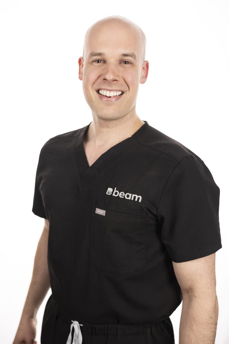
Dr. Clerk is a radiologist and fellowship-trained interventional radiologist with a wide array of experience in both interventional pain management and diagnostic imaging. In addition to providing expert patient care, Dr. Clerk places utmost importance on building a compassionate practice that recognizes patients as people, not numbers.
When you choose Beam, you can be confident that Dr. Clerk will stay with you throughout your care journey and help you make smart decisions about your pain and imaging needs.
Université de Sherbrooke
Medical School
Université de Sherbrooke
Residency | Diagnostic Radiology
Harvard Medical School
Fellowship | Neuroradiology
The Spine Fracture Institute
Fellowship | Interventional Pain Management