
The biopsy is a type of procedure that removes a small amount of tissue or fluid from a nodule to analyze its composition. Breast biopsy is performed under imaging guidance to ensure accuracy when taking a sample. The sample is then sent to a laboratory to be processed and characterized.
Breast biopsy is a medical procedure used to diagnose breast abnormalities or concerns, such as lumps or suspicious findings on a mammogram or ultrasound. It involves the removal of a small sample of breast tissue for examination under a microscope.
There are different types of breast biopsy procedures, including:
1. Fine-needle aspiration (FNA) biopsy: A very thin needle extracts fluid or cells from a lump or cyst in the breast.
2. Core needle biopsy: A slightly larger needle is used to remove a cylindrical sample, or core, of breast tissue, including both the interior and surrounding tissue.
Breast biopsies are typically performed under local anesthesia to minimize discomfort. The procedure may be guided by ultrasound, or mammography to ensure accurate and precise sampling of the suspicious area.
Once the tissue samples are obtained, they are sent to a pathologist who examines them under a microscope to determine if the cells are cancerous or benign. The results of the biopsy can help guide further treatment decisions and ensure appropriate management of the breast abnormality.
Overall, breast biopsy is an important procedure for diagnosing breast abnormalities and determining whether they are cancerous or non-cancerous. It allows for accurate and targeted examination of the breast tissue to provide a definitive diagnosis and inform an appropriate treatment plan.
Cyst aspiration describes using a very thin needle to puncture a cyst and remove the fluid within it, collapsing it. Cysts are non-cancerous and can be aspirated if they have gotten too large or are causing discomfort. The fluid from within can be sent for analysis if the radiologist feels that it is appropriate to do so.
Before the Procedure:
During the Procedure:
During an ultrasound-guided breast biopsy or cyst aspiration, you will lie on your back on an examination table. The radiologist or sonographer will apply a gel to your breast to improve the contact between the ultrasound probe and your skin. They will then use the ultrasound probe to locate the area of interest in your breast. This is called a pre-scan.
Once the area of interest is identified, the radiologist will numb the area with a local anesthetic (using a very fine needle) to minimize any discomfort. They will use the ultrasound to guide the needle into the area of interest. This helps the radiologist ensure accurate and precise targeting of the desired tissue. The needle will be used to remove tissue, fluid, or collect cells, and this sample will be sent to a laboratory to be analyzed.
You will be awake and can ask any questions you like during the biopsy. This is a straightforward and well-tolerated procedure that plays a crucial role in diagnosing breast diseases and guiding appropriate treatment decisions. The risk of complications is very low.
After the Procedure:
After the biopsy is completed, pressure will be applied to the biopsy site to minimize bleeding. A small bandage or dressing will be applied to cover the site.
You will be given specific instructions on how to care for the biopsy site, such as keeping it clean and dry, and avoiding strenuous activity (jogging) or heavy lifting for 1 week following the biopsy.
You may experience some discomfort or soreness at the biopsy site, which can be relieved with over-the-counter pain medications. It is normal to have some bruising and swelling, it can appear extensive but typically resolves within a few days. Apply ice to the area to decrease discomfort and help to minimize bruising, while avoiding heat applied directly to the area as this will increase bruising. Wearing a sports bra can provide support and comfort, as well as a way to hold the icepack against the breast.
The tissue sample collected during the biopsy will be sent to a laboratory for analysis. The results will be communicated to your healthcare provider, who will discuss them with you and determine the next steps based on the findings. If the biopsy reveals cancerous cells, further diagnostic tests and treatment options will be discussed with you.

If you have any questions or would like to learn more, please
contact us. We look forward to supporting your journey to better health.

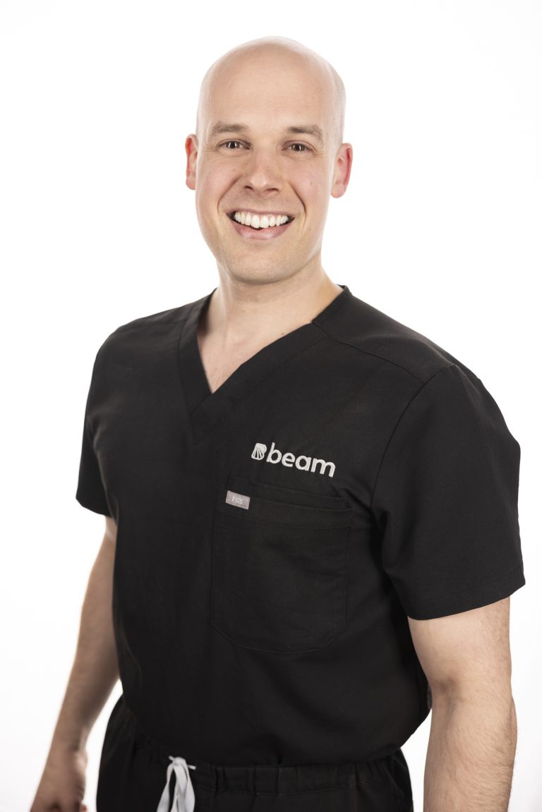
Dr. Clerk is a radiologist and fellowship-trained interventional radiologist with a wide array of experience in both interventional pain management and diagnostic imaging. In addition to providing expert patient care, Dr. Clerk places utmost importance on building a compassionate practice that recognizes patients as people, not numbers.
When you choose Beam, you can be confident that Dr. Clerk will stay with you throughout your care journey and help you make smart decisions about your pain and imaging needs.
Université de Sherbrooke
Medical School
Université de Sherbrooke
Residency | Diagnostic Radiology
Harvard Medical School
Fellowship | Neuroradiology
The Spine Fracture Institute
Fellowship | Interventional Pain Management