
Automated breast ultrasound, also known as ABUS, is a specialized imaging technique used in addition to mammography for breast cancer screening. It involves the use of an automated ultrasound device that produces detailed images of the entire breast using sound waves. Breast ultrasound does not use radiation.
ABUS is particularly useful in women with dense breast tissue, as dense tissue can make it more challenging to detect abnormalities or lesions on mammogram. In these cases, ABUS can provide additional information and improve the detection of breast cancer.
While ABUS can provide valuable information, it is important to note that it is not a replacement for mammography. Mammography remains the mainstay of breast cancer screening, and ABUS is used as an adjunct to mammography in specific cases. Women need to discuss their individual risk factors and screening recommendations with their healthcare provider to determine if ABUS is appropriate for them.
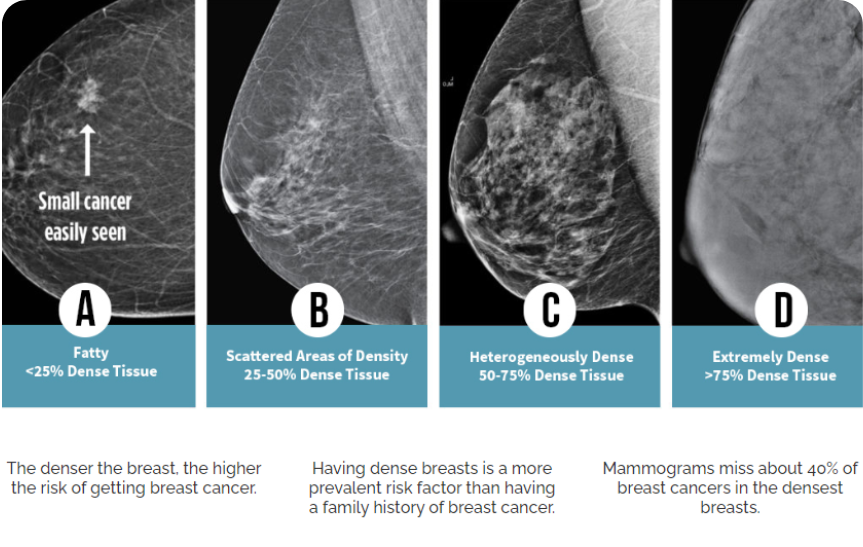
ABUS is particularly useful for women classified as having dense breast tissue on mammograms. This is not something that can be determined by simply looking at the breast.
When a person has dense breast tissue it makes the mammogram more difficult to read, and therefore less sensitive to small changes or abnormalities.
ABUS can work in conjunction with mammograms to provide a robust breast imaging protocol for dense breasts.

Used with permission from https://densebreastscanada.ca/
You will be asked to change into a gown from the waist up. The technologist will assist you in getting positioned with a pillow, lying down on the ultrasound bed. During an ABUS exam the ultrasound transducer will be using high frequency sound waves to generate images of the breast tissue from multiple angles. This is very similar to a classic handheld ultrasound experience. These images are then processed to create a three-dimensional image of the breast.
ABUS is a non-invasive and painless procedure that does not involve radiation. The exam typically takes about 15 to 30 minutes to complete, and the results are reviewed by a radiologist.
After the radiologist has received the images, they will review and compile a report for your referring care provider. This is typically received in 1-4 days from the time of the appointment. Included in the report for the referrer will be relevant findings and the corresponding recommendations according to the current guidelines.

If you have any questions or would like to learn more, please
contact us. We look forward to supporting your journey to better health.

Dr. Clerk is a radiologist and fellowship-trained interventional radiologist with a wide array of experience in both interventional pain management and diagnostic imaging. In addition to providing expert patient care, Dr. Clerk places utmost importance on building a compassionate practice that recognizes patients as people, not numbers.
When you choose Beam, you can be confident that Dr. Clerk will stay with you throughout your care journey and help you make smart decisions about your pain and imaging needs.
Université de Sherbrooke
Medical School
Université de Sherbrooke
Residency | Diagnostic Radiology
Harvard Medical School
Fellowship | Neuroradiology
The Spine Fracture Institute
Fellowship | Interventional Pain Management