CT - Computed Tomography Scan
What is a CT Scan?
Utilizing the lowest radiation dose possible, a three dimensional (3D) x-ray imaging machine, and computer software, CT scans are used to diagnose abnormalities in most of the areas of the body including the brain, spine, chest, abdomen, pelvis – including the organs within this area, bowel, bone, blood vessels, and soft tissues. In some instances, your test may require the use of an intravenous or oral contrast dye to be used to enhance vessels or organs to better visualize the area of interest. CT scans provide high-resolution images with greater detail than a standard x-ray.
The exam is performed by an x-ray technologist with special training in CT scanning and the images are analyzed and reported by a radiologist.
For patients with claustrophobia and/or anxiety
Pricing
$425 – Head
$550 – Chest
$425 – Bones/Joints/Spine
$725 – Neck/Sinus/Facial Bones
$850 – Abdomen/Pelvis
$125 – Contrast (if required)

Types of CT Scans
Our diagnostic tools and team of specialists provide Calgary and the surrounding areas with access to personalized service and care.

CT Head
A head CT scan is a painless, non-invasive procedure that uses x-rays to create detailed images of your head, including the skull, brain tissue, and blood vessels. A head CT can detect bleeding in the brain, tumors, fractures, head injury, or an aneurysm.
CT Spine
Provides a detailed assessment of the bones and joints that make up some of the spine including arthritic changes, fractures, and malalignment. CT scans provide some information of the soft tissues of the spine, but MRI may be more sensitive.

CT Extremities
A CT scan of the extremities include the shoulder, arm, elbow, wrist, hand, hip, leg, knee, ankle and foot. This scan provides detailed images of the anatomy of bones, including evaluating fractures, bone tumours, osteomyelitis, arthritis and other causes of chronic joint pain.
CT Chest
A CT scan of the chest helps to diagnose various lung problems, including blood clots, lung tumors or masses, fluid around the lungs, COPD, pneumonia, scarring of the lungs, tuberculosis or interstitial lung disease with scarring.

CT Abdomen
A CT scan of the abdomen evaluates and provides a more detailed visualization of organs including liver, stomach, spleen, pancreas, kidneys, colon, aorta and vital blood vessels.

CT Pelvis
A CT scan of the pelvis helps to diagnose conditions in the bladder, pelvic organs, cancers in the pelvic region, appendicitis and pelvic inflammatory diseases.
FAQ
- Depicts smaller hard to see fractures that Xray may not catch
- Cancer
- Disease
- Trauma
- Soft tissue injuries
- Head
- Spine
- Extremities
- Chest
- Heart
- Abdomen
- Pelvis
- Soft Tissue
- Lung Screening
- Colonography, also known as Virtual Colonoscopy
It is important you discuss pre-existing medical conditions and medications you are taking prior to your CT examination with your doctor. Special preparations may be required.
In general, you may be asked to change into a gown for your exam, remove any metal objects, and refrain from eating or drinking for a few hours prior to your exam.
Upon arrival, the CT scan process can be expected to take less than 30 minutes with the scan itself only occupying a few minutes.
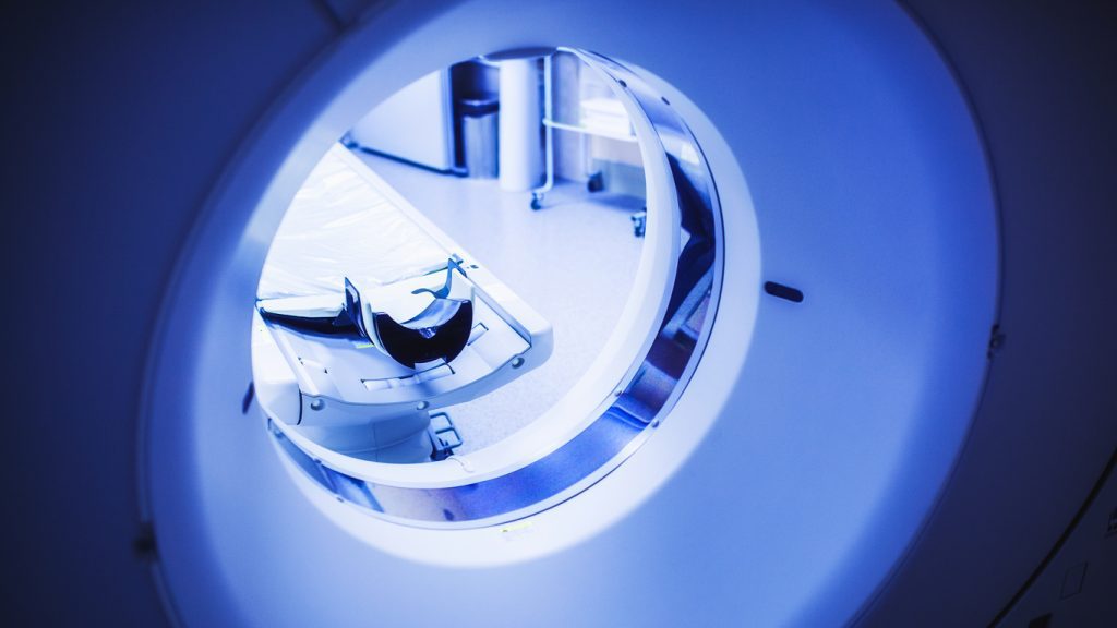
When you have reached the examination room, you will be positioned on the CT imaging table. This table will move you in and out of the CT machine as it obtains 3D imaging of the area of interest.
Throughout the scan the technologist will be able to communicate with you. They will be able to hear anything in the room, as well as monitor the scan, and necessary injection of contrast when this is indicated.
While you are within the scanner (CT machine) you will hear a humming as the machine performs the imaging.
CT contrast may be utilized depending on the ordered imaging. Patients who are of the age 60 and older that require contrast for their scan will be asked to provide up to date bloodwork (within 3 months) to ensure adequate kidney function.
During your CT scan you will be exposed to low levels of radiation. Although the level of radiation from CT scans is higher than those of an x-ray, CT scans have not been shown to cause long-term effects.
If you are pregnant or if you think you may be pregnant, it is important to discuss your CT exam with your doctor prior to your appointment. Although the radiation from a CT exam is not likely to cause any harm to your baby, your doctor may prescribe another diagnostic imaging procedure such as an MRI or Ultrasound.
If CT contrast is to be used during your procedure, it is important to discuss with your doctor prior to the exam. Patients that are allergic to contrast material may experience minor rash or itchiness, however, more extreme reactions are possible. If you are a patient with Renal Disease or Diabetes, it is also important to discuss the use of CT contrast in your exam.
Some patients who suffer from anxiety or claustrophobia may experience a level of discomfort while the exam is being performed. If you think this may be an issue for you, please let us know prior to your appointment. There are some options that may be available to you such as; minimal sedation (helps you to relax and change your mood – ex. Ativan) or conscious sedation (makes you feel sleepy, but you have full control of all reflexes – ex. Penthrox).
CT contrast is a special dye used in some CT exams to help highlight the areas of your body that are to be examined. The dye is designed to appear white on CT images, which helps emphasize internal structures of the body in CT images. It is important to discuss the use of CT contrast with your doctor prior to your procedure.

Additional CT Screening Options
Through The Beam Health Assessment Program
Elite CT Screening
The Elite Screen is a combination of the Total MRI Screen + LDCT Lung Screen + LDCT Colonography (Virtual Colonoscopy).
This screen focuses on your entire body by utilizing both MRI & CT.
Complete Scan
Get a complete scan of the lungs, colon/ large bowel and abdomen and pelvis.


Low Dose Lung & Colon Scan
Combine the low dose lung screen and low dose colonography into one.
Low Dose CT Colonography Screen
CT Colonography is a procedure that uses CT scans to look inside your large bowel. It’s used to check for colonic polyps, a pre-cursor to cancer, and to investigate bowel symptoms.


Low Dose CT Lung Screen
When it comes to lung cancer, earlier detection means better prognosis.
Interested in booking a CT screen?
Your Health is Important to Us
If you have any questions or would like to learn more, please
contact us. We look forward to supporting your journey to better health.

