Breast Health Program
Breast Imaging Basics
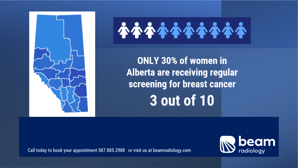
Regular mammograms can help save lives by identifying breast cancer at its earliest and most treatable stage, often long before it can be felt.
Screening mammography is used as a preventative tool to detect breast cancer at an early stage in women without symptoms or any known concerns. They are performed at routine intervals depending on age and other risk factors.
Diagnostic mammography is used to evaluate a specific concern or abnormality identified in the breast. This can be prompted by the patient or doctor noticing a change or abnormality in the breast, or as a follow up to a prior set of imaging.
In addition to mammography the breast health program at Beam Radiology also includes a variety of other services including automated breast ultrasound, diagnostic breast ultrasound, breast biopsy, and breast MRI.
Why screen?
75% of women who develop cancer have no family history of breast cancer.
Of those women dying of breast cancer, 75% had no regular screening, 15% had regular screening, and 10% had a malignancy not detected on mammography.
Radiation risk: In 100,000 patients, 200 will develop breast cancer. Fewer than 6 will develop a radiation related breast cancer – and the mammogram will likely detect that cancer.
In Alberta women aged 45 – 74 can self refer for screening mammogram. They do not need a referral or requisition.


Mammography
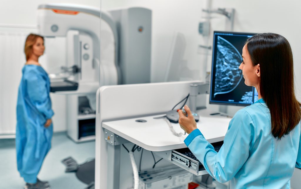
Mammography is a special type of medical imaging that is used to detect and diagnose breast diseases, including breast cancer. It involves taking low-dose X-ray images of the breasts to look for any abnormalities or changes in the breast tissue even before a patient or doctor can feel a lump. At Beam, we offer both screening and diagnostic mammograms.
Automated Whole Breast Ultrasound
Automated whole breast ultrasound, also known as ABUS, is a specialized imaging technique used in addition to mammography for breast cancer screening. It involves the use of an automated ultrasound device that produces detailed images of the entire breast using sound waves.
Breast ultrasound does not use radiation.


Diagnostic Breast Ultrasound
Diagnostic breast ultrasound is an ultrasound of the breast tissue used to investigate a specific concern, new breast symptom, or as a supplement to another screening exam such as mammography. The ultrasound provides imaging of the internal breast tissue by using a transducer and ultrasound gel, just like an ultrasound of a different part of the body.
This is a painless exam and does not use radiation.
Breast Intervention
There are various breast interventions performed at Beam including biopsy and aspiration.


Comprehensive Genetic Screening
Beam Radiology is committed to offering the best available options to our patients. Part of this is examining the emerging resources and the scientific innovations that we now have access to. This includes GENETIC SCREENING.
Knowledge is power. Know your health-related genetic risks. Insight into your health-related risks allows for the opportunity to be proactive.
Not only are you investing in your own health, but you are investing in theirs. With the genetic screening add-on, any positive result allows for your blood relatives to have the opportunity to also receive screening at NO COST.
Your Health is Important to Us
If you have any questions or would like to learn more, please
contact us. We look forward to supporting your journey to better health.

